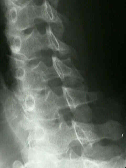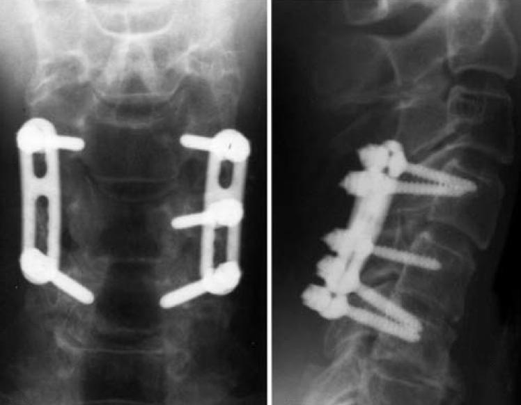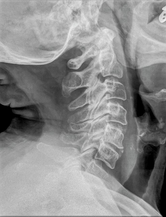

Normal in adults: 1 is suggestive of occipitocervical dislocation.


McGregor’s line: From superior margin of hard palate to inferior margin of occiput abnormal if odontoid tip >4.5 mm above this line.Chamberlain’s line: Line drawn from posterior margin of foramen magnum (opisthion) to superior margin of hard palate abnormal if tip of odontoid >3 mm above this line.McRae’s line: Line drawn from anterior and posterior margin of foramen magnum (basion to opisthion) difficult to evaluate in X-ray.Odontoid view may show odontoid fractures which can be classified by Anderson and D’Alonzo classification:.Bow tie sign (Unilateral facet dislocation): bodies appear true lateral below level of injury and oblique above level of injury.Forward shift of vertebral body: 25% suggests unilateral facet dislocation and 50% suggests bilateral facet dislocation.Flexion views: >11 degrees angulation using end-plate method and >3.5 mm translation between posterior vertebral lines (anterior translation) is suggestive of mechanical instability.Harris ring: Sclerotic ring formed inside body of C2 by it’s lateral masses (break in the ring outline is suggestive of fracture of peg/body of C2).Idenitfy “Coffee Bean” shadow – anterior arch of C1 vertebra always visible on lateral view.Trace outline of each pedicle, lateral mass, lamina and spinous process.Crushed vertebral body (burst fracture) or flexion deformity with antero-inferior tear drop (tear drop fracture) are unstable.Trace outline of each vertebral body – below C2 they must be approximately same height and size.Line 3: Make sure there is no asymmetry of the articular spaces between the dens and the lateral masses of C1.Line 2: Make sure there is no asymmetry of the articular spaces between the lateral masses of C1 and the body of C2.Line 1: Make sure lateral masses of C1 doesn’t hang over the lateral masses of C2 ( rule of Spence: If combined overhang of lateral masses of C1 over C2 on both sides is >7 mm – transverse ligament injury making Jefferson or C1 ring fracture unstable).Two lateral lines connecting lateral masses.Spinous process line (must be in midline).A normal dens tilts posteriorly and, if it appears straight or anteriorly tilted, fracture should be considered.Atlanto-occipital alignment: Anterior margin of foramen magnum should line up with dens (clivus point at dens) and Posterior margin of foramen magnum should line up with C1 spino-laminar line.There are lines that must run uninterrupted – 3 views: True lateral, AP, Odontoid (Open mouth) +/- Swimmer’s view (when a standard lateral view cannot image the cervicothoracic junction).Skull base, C1-C7 and upper T1 must be visible.Lamiot, CC BY-SA 3.0, via Wikimedia Commonsįor images of the particular Cervical spine (C-spine) X-ray findings and views mentioned below, please refer to the links at the end of the article or use web-search. Cervical_Xray_Extension.jpg: StillwaterisingCervical_Xray_Extension_view.jpg: Stillwaterisingderivative work: F.


 0 kommentar(er)
